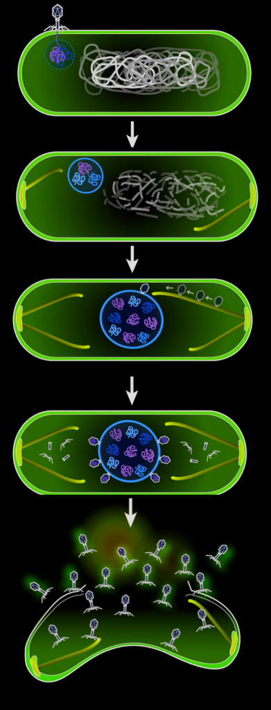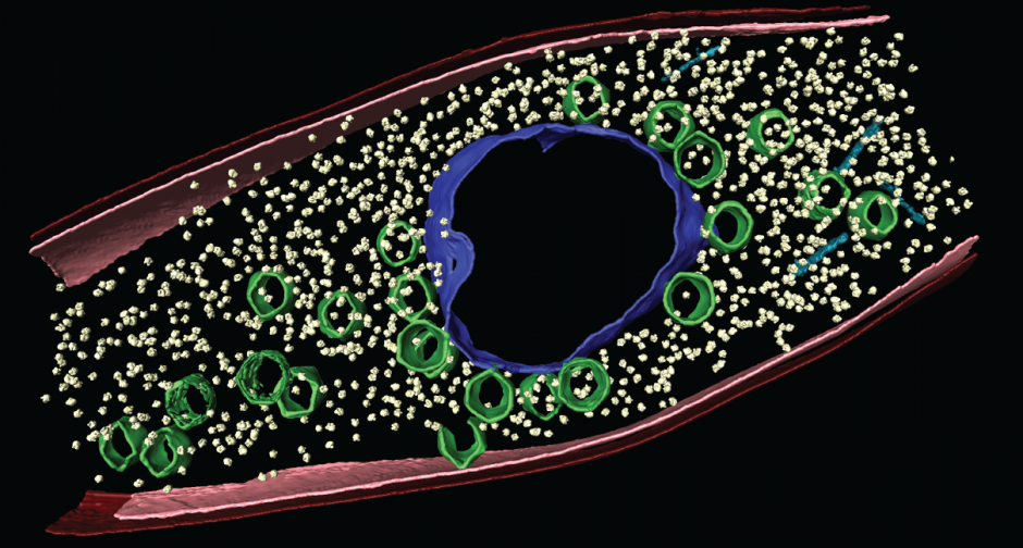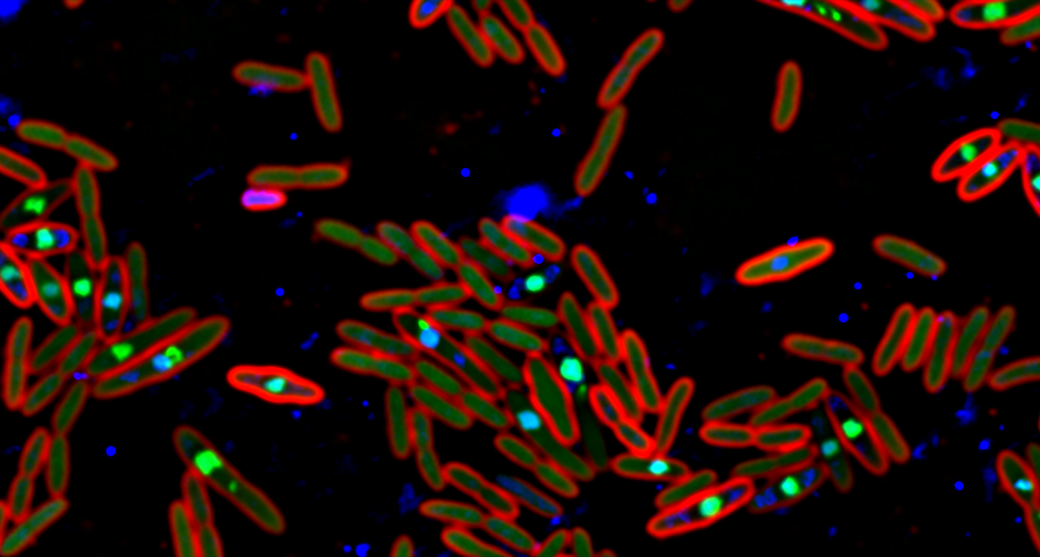
The nucleus is the cellular structure that defines Eukaryotes, separating them from Bacteria and Archaea. We recently described the first example of a nucleus-like structure that is assembled in bacteria upon infection by a virus. During lytic growth, many Pseudomonas jumbo phage including PA3, PhiKZ, and 201φ2-1, build a compartment that physically separates viral DNA from the cytoplasm. The “phage nucleus” assembles immediately after DNA injection, grows larger as the DNA replicates, and is positioned at midcell by a bipolar tubulin-based spindle. Protein localization data show that DNA replication and transcription occur inside the phage nucleus, while translation and metabolic processes, such as nucleotide synthesis, occur outside of the nucleus. Time-lapse microscopy demonstrates that proteins synthesized in the cytoplasm later accumulate inside the phage nucleus, suggesting a two-way exchange of mRNA, protein, and nucleotides between the cytoplasm and nucleus, as in Eukaryotes. Late in infection, viral capsids assemble on the cell membrane and traffic through the cell along tread-milling PhuZ filaments. Capsid dock on the surface of the phage nucleus, where DNA packaging occurs. Ultimately, fully assembled viral particles form and are released when the cell lyses. These results demonstrate that bacteriophage evolved both a spindle and a nucleus, providing insight into the evolution of these defining structures of eukaryotic cells.
10
Jun
Postdoctoral positions available
Postdoctoral positions available. If you are interested in the wonderful and bizarre world of bacterial cell biology, and what their giant phage foes make them do, join us!

